This section summarises the different treatment options available for intraocular and extraocular tumours. After leaving clinic, you may have forgot to ask a question about the treatment advised. This is very common when being faced with a diagnosis of eye cancer. We hope this section will help answer your questions. Please see the different treatment options below.
Intraocular Tumour Treatments
Below is the list of treatments available to us for tumours that grow inside the eye. The most common tumour that grows inside the eye is melanoma. The most common treatment for this is plaque radiotherapy. If the tumour is too big for this treatment we may consider proton beam radiotherapy. We aim to select the best treatment suited to each patient as an individual. Unfortunately this sometimes means having to remove the eye (enucleation). Please see the different treatment options below.
Plaque Radiotherapy
Plaque radiotherapy is a form of internal radiotherapy. A radioactive piece of metal known as a plaque is attached to the sclera (white part of the eye) next to the tumour. This is around the size of a 5 pence coin (please see picture above). This is done in the operating theatre and is left in the eye between 3 and 7 days before being removed. Patients usually stay in ward 1C for this treatment. The tumour starts to shrink around 4-6 months after the plaque is removed.
The effects can last for several years. Although being an effective treatment, the radiation can sometimes damage other parts of the eye. This may cause cataract, retinal detachment, nerve damage, or macular oedema (swelling of the back of the eye). New blood vessels may grow after treatment; these can sometimes block the drainage angle in the eye causing glaucoma. If we are unable to control this, we may have to consider removing the eye. Although there are risks of plaque radiotherapy, this treatment can stop growth of the tumour in around 80% of cases.
Proton Beam Radiotherapy
This is where radiation with charged particles called protons are targeted at the tumour from outside the eye. This treatment is used if the tumour is too large or located too far back in the eye for plaque radiotherapy to work. As this treatment is highly specialised, the Douglas Cyclotron Unit, Clatterbridge, Liverpool is the only centre in the United Kingdom where the treatment is given.
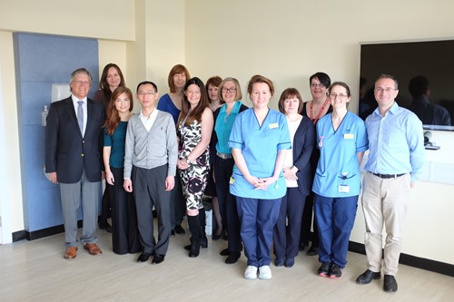
If proton beam radiotherapy is the best option for you, we will organise transport to Clatterbridge and accommodation for you in near the Hospital (please see the team photo of the Clatterbridge team and the hospital above). This involves two separate visits to the Clatterbridge. At the first visit a treatment mask is made and fitted- this helps the team target the radiotherapy at the correct part of the eye. At the second visit, one to two weeks later, the treatment is given. You will likely travel down Sunday evening and return Friday later that week. Final measurements are made on the Monday and then the radiation is given over the remaining four days during (Tuesday to Friday).
Each treatment session takes around 20 minutes and is pain free. Before going down to Liverpool, however, we have to perform a small operation on the affected eye in Gartnavel General Hosptial, Glasgow. This is where we stitch tantalum markers (small metal discs smaller than a paper clip- please see photo above) to the sclera (white part of the eye) next to the tumour. We usually do this under General Anaesthetic. This helps the team target the radiation treatment more effectively when you go down to Clatterbridge. Proton beam radiotherapy takes a little longer to work than plaque radiotherapy.
We usually wait six months to see if the tumour starts to decrease in size. Although effective at treating tumours, the radiation can also damage normal parts of the eye and tissues around the eye when given. This may cause loss of eyelashes, loss of pigmentation of the eyelids, and inflammation of the conjunctiva causing a watery eye. Sometimes small blood vessels can grow at the back of the eye and into the drainage angle at the front of the eye. This can cause the pressure to build up in the eye and cause glaucoma. If we are unable to control the pressure and the eye becomes very uncomfortable, unfortunately we may have to consider removing the eye.
External Beam Radiotherapy
This is where radiation from a machine is targeted at cancer cells from outside the body. We find this treatment useful in patients that have cancer else where in the body that has spread to the back of the eye. On the other hand, if there has been a large tumour inside the eye that has grown behind the eye, we may choose to use radiotherapy after removing the eye. This treatment is spit into fractions.
This means the treatment is divided into smaller doses and spread out over a few days. This gives healthy cells in the body a chance to recover between treatments. If we are using radiation to treat the eyes or lymph nodes around the head and neck, then a special mask is usually made for patients. This mask is worn during treatment and helps aim the radiation at only the cancer cells. Although this helps prevent damage to healthy cells around the tumour, treatment may still cause loss of eyelashes, loss of pigmentation of the eyelids, and inflammation of the conjunctiva causing a watery eye.
Laser Transpupillary Thermotherapy
Transpupillary Thermotherapy (TTT) uses an infrared laser beam to heat the tumour up and kill the cancer cells. This technique is useful if there is uncertainty if the suspicious area is a melanoma or a naevus, or if the choroidal melanoma is small and radiotherapy is inappropriate due to poor health.
This treatment is sometimes combined with plaque radiotherapy as it reduces swelling and leakage from the blood vessels. After TTT the tumour gradually shrinks down if successful. Repeat treatment may be required at 6 months.
Possible complications from this treatment include retinal detachment, blockage of blood vessels, growth of new blood vessels, iris burns and cataract. Unfortunately tumour recurrence after treatment is common. This is more likely to occur if the melanoma is thick, close to the optic nerve, or non-pigmented.
Photo Dynamic Therapy
A light sensitive dye is injected into the blood stream. As the dye travels through the blood vessels to the back of the eye through the blood vessels, a special light is shined into the eye. This activates the dye and causes the abnormal blood vessels to close, shrink, and stop leaking.
This is useful in patients with a naevus or melanoma that is leaking fluid and building up at the back of the eye (macular oedema). Although this is not the main treatment for choroidal melanoma, we may find it useful in some cases where radiotherapy treatment is not possible.
Removal of Eye: Enucleation
Enucleation is the medical term for removing the eye. This is recommended when the tumour is too large for other treatments or has started to invade behind the eye. Removing the eye along with the tumour is sometimes preferred if there is a lot of pain and discomfort in the eye. This can be due to high pressure inside the eye caused by blockage of the drainage angle by tumour or new blood vessels grown after radiotherapy.
The idea of having your eye removed is scary. With current technology, however, we can get excellent cosmetic results with uniquely designed and fitted artificial eye implants (please click below to see photos). Our artifical eye clinic is run in Gartnavel General Hospital, Glasgow. Alternatively, some patients may prefer to just wear an eye patch after the tumour is removed.
After removing the eye, although this gets rid of the tumour growing in the eye, unfortunately this does not prevent the tumour growing else where in the body later in life. The most likely place for melanoma to regrow is the liver. For this reason we may decide to organise a liver ultrasound scan every year at your local hospital to screen for cancer growth.
Artificial Eyes
After the eye is removed, a temporary cosmetic shell is fitted in the operating theatre (see first picture above). We choose a colour to match the patient’s other eye. Although not a perfect colour match, this temporary shell will remain in place until the final artificial eye is made.
Once the eye socket has healed, the temporary cosmetic shell is removed and the final artificial eye implant can be fitted. This is done in the prosthetic eye department in Gartnavel General Hospital, Glasgow. The team will take photos of your normal eye. The artificial implant is then painted in fine detail to match the photo of your healthy eye. Please see the two other photos above (note both the healthy eyes have been dilated in clinic to examine the back of the eyes so the pupil sizes do not look symmetrical).
Removal of Eye and contents of Eye Socket- Exenteration
Exenteration means removing the eye with the tumour and the soft tissue around the eye. This treatment is required if the cancer has spread behind or around the eye. Sometimes the eyelids or part of the bone around the eye have to be removed if the tumour has invaded here. If this is the case, we will likely perform the operation at the Queen Elizabeth University Hospital with the help of our Oral and Maxilofacial Surgeon colleagues. Sometimes this treatment is combined with radiotherapy or chemotherapy. Our medical oncology team will help us choose the best treatment for you.
Excellent cosmetic results can be achieved after the tumour is removed. This involves further reconstructive surgery and being fitted with an artificial eye. Some patients, however, may prefer just to wear an eye patch or leave things as they are instead of having further surgery. After the cancer is removed we can plan treatment that suits your needs.
Extraocular Tumour Treatments
Please see below the list of treatment options for tumours that grow outside the eye. Every case is different. In clinic we will discuss the best treatment or combination of treatments for you.
Surgical Excision of Eyelid Tumours
This can be performed under local or general anaesthetic and as a day case. After tumour removal as much normal tissue is left behind to help keep the eyelid looking as normal as possible. The tumour is sent to the pathology laboratory to confirm the type of tumour and if it has all been removed. Eyelid tumours (mainly basal cell carcinomas) may be removed by a dermatologist (skin specialist). This is where a small part of the skin is removed then inspected under a microscope straight away. If cancer cells are still visible, then more tissue is removed and inspected again. This is repeated until there are no more cancer cells seen under the microscope. This helps remove as little normal tissue as possible mean while ensuring all the cancer is removed.
After the tumour is removed the eyelid is reconstructed to get the eyelid looking and functioning as normal as possible. If the tumour removed was small then this can usually be done on the same day. If the tumour removed was large and a lot of the eyelid had to be removed then reconstruction may be done on a different day. Skin or tissue can be taken from the other eyelid or from other parts of the body to re-form the eyelid. We commonly use the skin in front of the ear or from the inner surface of the upper arm. Sometimes we use tissue from the inner surface of the cheek- this heals very well after surgery. As there are many options, we aim to choose the best treatment option for you.
Freezing treatment: Cryotherapy
This freezes the tumour helping destroy the cancer cells. This can be used in combination with surgical excision, or on its own if surgery is not an option. Cryotherapy can is usually done the operating theatre under local or general anaesthetic. Treatment lasts several minutes. Although helping prevent tumour growth, no samples are sent to the lab so confirmation of tumour death is not always possible. If this is the case, we will monitor you carefully in the clinic. Sometimes we have to repeat this treatment more than once.
Radiotherapy
This treatment involves targeting the cancer with high energy radiation beams. This kills the cancer cells and stops them multiplying. This is used if surgery is not possible, for example, if the patient is too unwell or desperately does not want surgery. It may, however, be the preferred treatment of choice; for example, in lymphoma. This treatment is performed as an out-patient. This treatment is spit into fractions. This means the treatment is divided into smaller doses and spread out over a few days. This gives healthy cells in the body a chance to recover between treatments. If we are using radiation to treat the eyes or lymph nodes around the head and neck, then a special mask is usually made for patients. This mask is worn during treatment and helps aim the radiation at only the cancer cells. Although this helps prevent damage to healthy cells around the tumour, treatment may still cause loss of eyelashes, loss of pigmentation of the eyelids, and inflammation of the conjunctiva causing a watery eye.
Chemotherapy
Mitomycin C (MMC)
This is a chemotherapy drug that is applied to the surface of the tumour in theatre. MMC works by sticking the cancer cells’ DNA (the cell’s genetic code) together, stopping the tumour or cancer cells from growing.
This is applied to the surface of the cancer cells and therefore side effects of chemotherapy such as nausea, vomiting or hair loss are not experienced.
If MMC eye drops are being used, however, this can irritate the eye. We may give lubricant or steroid drops to treat this.
5-fluorouracil (5-FU)
This treatment, also called imiquimod, is a chemotherapy drug which is applied to the surface of skin tumours. Sometimes we use this to treat eyelid tumours. This drug causes the body’s immune system to produce a chemical called interferon. This attacks and kills cancer cells. It may irritate the skin when applied, this means the treatment is working. It is applied 3 – 5 nights a week and treatment can last up to 6 weeks.
Our service is located in the:
Ophthalmology Out Patient Department Gartnavel General Hospital 1053 Great Western Road, Glasgow G12 OYN
Please click on “travel” below to help plan your journey. If you plan to travel the day before the clinic, please click on “accommodation” below.
Travel
We have bus and train links that travel to Gartnavel General Hospital daily.
Traveling by Train
Hyndland Station is the nearest train stop to Gartnavel General Hospital. We are a 5 to 10 minute walk from the Hyndland Station. Trains leave both Glasgow Queen Street Station and Glasgow Central Station every 10-15 minutes. The journey takes roughly 10 minutes to get to there. The following websites will help you plan your train journey if you are traveling from else where in Scotland:
Buses run frequently to from Glasgow City Centre to Great Western Road; this is a 5 minute walk to the main entrance to the hospital. Travel time may take up to 30 minutes during rush hour. The buses run every 10-15 minutes from the city centre. Please see the following links for time tables and to plan your journey:
When visiting family members on the ward, people who are elderly or live with a disability may find the evening hospital visitor transport service useful:
Parking is extremely limited at Gartnavel General Hospital. A maximum four hour stay operates in patient and visitor car parks from 7.30am until 4.30pm Monday to Friday. If there are no spaces, the Glasgow Pond Hotel next to Gartnavel General Hospital offer parking at reasonable rates. There is a designated drop off points outside all the entrances to the hospital. Please avoid parking on disabled spaces unless you hold a valid badge, and also taking care not to park on ambulance car parks or yellow lines.
Accommodation
We appreciate that patients and relatives may be travelling a long distance to attend the clinic. If you choose to travel the day before, Leonardos Inn Hotel is next to Gartnavel General Hospital. Please see their website for reservation and bookings.
The Scottish Ocular Oncology Service is run by Dr Cauchi, Dr Chadha and Dr Connolly, experienced consultant ophthalmologists with a specialist interest in ocular oncology. Over the years we have a built up a close team of doctors , nurses, and non-medical staff from different backgrounds. These include:
Ophthalmologists (Eye doctors, both at consultant and registrar level)
Radiologists (Experts in CT, MRI, and Ultrasound scans),
Oncologists (Cancer specialists),
Pathologists (Experts in analysing tumours)
Specialist ophthalmic nurses (Nurses trained in counselling and able to answer questions about your diagnosis and treatment).
Anaesthetists (Experts at putting you to sleep for your operation)
Service Coordinators
Medical Photographers
Every Thursday morning we have our multidisciplinary team meeting (MDT). This is where we discuss patients who were listed for treatment the week before, and new patients coming to the clinic that day. Below are synopsis of the doctors and nurses from our team.
Ophthalmologists
Dr Cauchi
Dr Cauchi graduated from the Royal Free Hospital, University of London in 1996. His first interest in ophthalmology developed as a medical student, following in the footsteps of his grandfather who was also an ophthalmologist. He then did extra training in oculoplastics, orbits and ocular oncology. Dr Cauchi is one of the consultant ophthalmologists who run the Scottish Ocular Oncology Service.
Dr Chadha
Dr Chadha graduated from the University of Delhi in 1997 and underwent his basic and higher specialist training in ophthalmology at Edinburgh before doing a Fellowship in Ophthalmic Oncology and Oculoplastic Surgery at Glasgow in 2008-2009. He has been a Consultant in the West of Scotland since 2009 and is now one of the Consultants responsible for delivering the Scottish Ocular Oncology Service.
Dr Connolly
Dr. Julie Connolly started her career in academic research, completing her PhD from the University of Glasgow before undertaking further research roles in the Beatson Institute for Cancer Research. She subsequently graduated from University of Glasgow medical school before completing Ophthalmology specialty training and a fellowship in ocular oncology and oculoplastics in the West of Scotland Deanery. Dr Connolly is one of the consultants responsible for delivering the Scottish Ocular Oncology Service.
Oncologists
Dr Ritchie and Dr Schipani are the two Consultant Oncologists that work with the Scottish Ocular Oncology Service.
Dr. Schipani
Dr Schipani graduated from the University of Milan (Italy) in 2001. He underwent his Clinical oncology specialist training from 2002 to 2006, University of Milano-Bicocca (Italy) and worked as a consultant oncologist in Italy for three years. After taking a consultant job in Glasgow in 2009, Dr Schipani has developed a specialist interest in treating eye cancers and joined the Scottish Ocular Oncology Service team since September 2016.
Dr. Ritchie
Dr Ritchie after receiving her medical degree from Glasgow University started her clinical oncology training in 1986. She became a consultant oncologist in 1993 and has a specialist interest in radiotherapy treatment for eye cancers and skin tumours around the eye. She is one of the two consultant oncologists that help decide the correct treatment for patients with eye cancer.
Radiologists
Dr Cram is a consultant radiologist who has a specialist interest in Ocular Radiology. Below is the background of his career to date.
Dr Cram
Dr Cram graduated from St Andrews University in 2003 and received his medical degree from Manchester University in 2006. He decided to become at radiologist in 2007 and studied radiology in the West of Scotland Deanery. After becoming a full time consultant in August 2013 he is now one of the two Radiologists who work with the Scottish Ocular Oncology Service.
Pathologists
Dr Roberts and Dr Thum are both consultant pathologists that work with the Scottish Ocular Oncology Service. A summary of their experience to date is outlined below
Dr Roberts
Dr Roberts graduated from the University of Glasgow in 1991. During her training in general pathology she undertook a fellowship at the University of Illinois at Chicago undertaking research for her MD in ocular toxoplasmosis. On returning to Glasgow she completed her training in ophthalmic pathology under Professor William Lee before taking up a consultant position in 1998. She is a member and former secretary and president of the British Association of Ophthalmic Pathology and a member and former secretary of the European Ophthalmic Pathology society. In conjunction with Dr Thum she provides eye pathology input for the Scottish Ocular Oncology Service.
Dr Thum
Dr Thum graduated from medicine in Aberdeen in 2001. After years of practising ophthalmology, he decided to pursue a career in Pathology and started his training in Edinburgh in 2007. He became a consultant pathologist in 2015 and has been working with the Scottish Ocular Oncology Service for one year. Dr Thum is one of the two pathologists in our team who examine cells and tissue from tumour samples to help us select the best cancer treatment.
Nurses
Our team of nurses play an integral role in counselling and caring for patients throughout their diagnosis and treatment. Below is a summary of Agnes, Julie, Gayle and Nichola’s experience and training to date.
Agnes Macleod
Charge nurse Macleod trained in the Western Infirmary, receiving her nursing degree in 1989. She has been working with the Scottish Ocular Oncology Service team since 1994. Having had completed the Professional Studies Ophthalmic and Counseling Skills courses, she provides care and support to eye cancer patients attending the clinic and staying on the ward.
Gayle Williamson
Staff nurse Gayle Williamson trained in Stirling University and graduated from nursing in 2012. She has completed her post-graduate Eye course in 2018 and her counselling course in 2019. She has been part of the Scottish Ocular Oncology team since 2016 helping provide care and support to eye cancer patients attending the clinic.
Nichola Campbell
Staff nurse Nichola Campbell trained in Glasgow Caledonian University and graduated in 2010. She began working in the ophthalmology ward in 2012 and has been working with the ocular oncology team since 2014. She plays an integral role in seeing patients through their initial diagnosis, treatment, and post-operative care.
Fiona Wallace
Staff nurse Fiona Wallace trained in Edinburgh Napier University and graduated from nursing in 2003, she has completed her post-graduate Eye course in 2021. She has been part of the Scottish Ocular Oncology team since 2008 helping provide care and support to eye cancer patients attending the clinic.
Scientists
More information coming soon….
Service Coordinator
Susan Ewan
Susan Ewan is the service coordinator for the Scottish Ocular Oncology Service. She has been providing comprehensive secretarial and administrative support to the Scottish Ocular Oncology Service since June 2004. Susan is the main point of contact for Health Professionals and patients alike. She arranges new appointments, scans and any treatments that may be required along with travel arrangements and transfer of information to the Douglas Cycloton Unit for patients having proton beam therapy.
For referrals please address letter to Dr Cauchi, Dr Chadha or Dr. Connolly at the following address:
Scottish Ocular Oncology Service Ophthalmology Out Patient Department Gartnavel General Hospital 1053 Great Western Road Glasgow G12 OYN
National Services Division Scottish Ocular Oncology Service
These guidelines are not intended to be prescriptive but to act as an aid to considering referral of patients to the nationally designated ocular oncology centre in Scotland at Gartnavel General Hospital. Glasgow.
Referring ophthalmologists should continue to exercise discretion based on the individual clinical presentations of their patients.
1. Whom to refer to the service
1.1 Patients with intraocular tumours
Any primary intraocular tumour other than naevus
Any intraocular metastatic tumour if specialist ocular oncology is required
Suspected intraocular lymphoma.
1.2 Patients with conjunctival and epibulbar tumours that appear invasive
1.3 Refer patient with conjunctival melanocytic tumour if :
Diameter exceeds 3 mm, especially in absence of clear cysts.
1.4 Patients with a suspicious melanocytic choroidal tumour having:
A: One of the following:
Thickness greater than 2.0 mm
Collar-stud configuration
Documented growth
B: Two of the following:
Thickness > 1.5mm
Orange pigment
Serous retinal detachment
Symptoms.
1.5 Refer patient with an iris nodule if:
Tumour is more than 3.0 mm in diameter
Tumour is markedly elevated
Secondary glaucoma or cataract
Tumour involves angle.
1.6 Patients with adnexal and orbital tumours if:
Eyelid tumour where ulceration and lash loss are evident or recurrence has occurred
Orbital tumours.
2. Whom not to refer to the service
2.1 Congenital hypertrophy of retinal pigment epithelium
2.2 Simple naevi, if:
Small and flat, or
Minimally raised with only drusen on the surface
Referral Form for Ophthalmologists
Please complete the referral form below and send it along with your referral letter. We would be grateful if these could be emailed along with any images, OCT or ultrasound scans.
Below are a list of publications produced by our department. We aim to continue producing good quality research to improve patient care.
Shams F, Cauchi P. Lagophthalmos and Ptosis in Inclusion Body Myositis. Ophthal Plast Reconstr Surg. 2016 Jan 18. [Epub ahead of print] PubMed PMID: 26784549.
Chia SN, Smith HB, Kemp EG. Comment on: ‘Pars plana vitrectomy to repair retinal detachment following brachytherapy for uveal melanoma’. Br J Ophthalmol. 2014 Apr;98(4):571. doi: 10.1136/bjophthalmol-2013-304749. PubMed PMID: 24390168.
Jamison A, Gregory ME, Lyall DA, Kemp EG. Visual outcomes following orbital biopsy. Orbit. 2013 Oct;32(5):304-8. doi: 10.3109/01676830.2013.814688. PubMed PMID: 23895509.
Cloke A, Lim LT, Kumarasamy M, Roberts F, Kemp EG. Lymphomatoid papulosis of the eyelid. Semin Ophthalmol. 2013 Jan;28(1):1-3. doi: 10.3109/08820538.2012.680643. PubMed PMID: 23305430.
Galea M, Cauchi P, Kemp E. Diode laser thermotherapy for conjunctival vascular malformations. Clin Exp Ophthalmol. 2013 Apr;41(3):307-8. doi: 10.1111/j.1442-9071.2012.02878.x. PubMed PMID: 22957708.
Irvine F, Kumarasamy M, Kemp E, Roberts F. Progression of primary acquired melanosis with atypia during pregnancy. Arch Ophthalmol. 2012 Aug;130(8):1085-7. doi: 10.1001/archophthalmol.2012.422. PubMed PMID: 22893092.
Achtsidis V, Gregory ME, Roberts F, Kemp EG. Enophthalmos following orbital trauma: a diagnostic catch. Br J Ophthalmol. 2012 Sep;96(9):1268-9, 1277. doi: 10.1136/bjophthalmol-2012-301996. PubMed PMID: 22872674.
Obi EE, Drummond SR, Kemp EG, Roberts F. Pleomorphic adenomas of the lower eyelid: a case series. Ophthal Plast Reconstr Surg. 2013 Jan-Feb;29(1):e14-7. doi: 10.1097/IOP.0b013e31825b34c1. PubMed PMID: 22743699.
Galea M, Falzon K, Chadha V, Williams G. Presumed occult globe rupture resulting in sympathetic ophthalmia. J Ophthalmic Inflamm Infect. 2012 Sep;2(3):137-40. doi: 10.1007/s12348-011-0056-4. PubMed PMID: 22200914; PubMed Central PMCID: PMC3438300.
Li Yim JF, Sandinha T, Kerr JM, Ritchie D, Kemp EG. Low dose orbital radiotherapy for thyroid eye disease. Orbit. 2011 Dec;30(6):269-74. doi: 10.3109/01676830.2011.615455. PubMed PMID: 22132844.
Wong KK, Roberts F, Cauchi P, Diaper C. Caliber persistent artery of the eyelid. Graefes Arch Clin Exp Ophthalmol. 2011 Sep;249(9):1395-7. doi: 10.1007/s00417-011-1685-x. PubMed PMID: 21494872.
Livingstone I, Ramamurthi S, Drummond S, Kemp E, Roberts F. Corneal perforation due to limbal involvement in Sézary syndrome. Graefes Arch Clin Exp Ophthalmol. 2011 Jul;249(7):1091-4. doi: 10.1007/s00417-010-1611-7. PubMed PMID: 21253759.
Macdonald EC, Cauchi P, Kemp EG. Proton beam therapy for the treatment of uveal melanoma in Scotland. Br J Ophthalmol. 2011 Dec;95(12):1691-5. doi: 10.1136/bjo.2010.195594. PubMed PMID: 21216794.
Obi EE, McDonald A, Kemp E. A bilateral cicatricial ectropion and bilateral upper lid shortening caused by 5-fluorouracil toxicity in a patient with dihydropyrimidine dehydrogenase deficiency. Cutan Ocul Toxicol. 2011 Jun;30(2):157-9. doi: 10.3109/15569527.2010.532846. PubMed PMID: 21077799.
Lim LT, Agarwal PK, Cauchi P, Diaper CJ. Laterality of periocular basal cell carcinomas in relation to driving practices in Scotland, United kingdom. Ophthal Plast Reconstr Surg. 2011 Jul-Aug;27(4):306. doi: 10.1097/IOP.0b013e3181f9e04b. PubMed PMID: 21057343.
Gregory ME, Chadha V, Roberts F, Kemp EG, Cauchi PA. Bilateral central retinal artery occlusion in a patient with primary central nervous system lymphoma. Graefes Arch Clin Exp Ophthalmol. 2011 Aug;249(8):1269-70. doi: 10.1007/s00417-010-1541-4. PubMed PMID: 20963435.
Lockington D, Chadha V, Russell H, Cauchi P, Tetley L, Roberts F, Kemp E. Histological evidence of tissue reaction to gold weights used for mechanical ptosis. Arch Ophthalmol. 2010 Oct;128(10):1379-80. doi: 10.1001/archophthalmol.2010.235. PubMed PMID: 20938017.
Lockington D, Chadha V, Russell H, Young D, Cauchi P, Kemp E. Socioeconomic status and choroidal melanoma in Scotland. Arch Ophthalmol. 2010 Mar;128(3):383-4. doi: 10.1001/archophthalmol.2009.407. PubMed PMID: 20212216.
Gonzalez P, Kemp EG, Roberts F. Recurrent choroidal melanoma after transscleral local resection with diffuse vitreous seeding. Graefes Arch Clin Exp Ophthalmol. 2010 May;248(5):741-6. doi: 10.1007/s00417-009-1261-9. PubMed PMID: 20127353.
Drummond SR, Kemp EG. Successful medical treatment of blepharochalasis: a case series. Orbit. 2009;28(5):313-6. doi: 10.3109/01676830903071190. PubMed PMID: 19874128.
Aziz S, Taylor A, McConnachie A, Kacperek A, Kemp E. Proton beam radiotherapy in the management of uveal melanoma: Clinical experience in Scotland. Clin Ophthalmol. 2009;3:49-55. PubMed PMID: 19668544; PubMed Central PMCID: PMC2708985.
Borooah S, Chadha V, Sutherland S. A case of permanent retinal disturbance postpartum following administration of ergometrine. Can J Ophthalmol. 2008 Oct;43(5):607-8. doi: 10.3129/i08-096. PubMed PMID: 18982049.
Ross JJ, Dean SJ, Koppel DA, Roberts F, Kemp EG. Massive orbital recurrence of uveal melanoma without metastases after 28 years. Br J Ophthalmol. 2010 May;94(5):632. doi: 10.1136/bjo.2008.146340. PubMed PMID: 18971235.
Ross JJ, Kemp EG. Large choroidal osteoma with macular decalcification. Retina. 2009 Mar;29(3):413-4. doi: 10.1097/IAE.0b013e3181871c2a. PubMed PMID: 18784621.
Chadha V, Cruickshank I, Swingler R, Sanders R. Advanced glaucomatous visual loss and oral steroids. BMJ. 2008 Aug 1;337:a670. doi: 10.1136/bmj.a670. PubMed PMID: 18676441.
Cauchi PA, Ang GS, Azuara-Blanco A, Burr JM. A systematic literature review of surgical interventions for limbal stem cell deficiency in humans. Am J Ophthalmol. 2008 Aug;146(2):251-259. doi: 10.1016/j.ajo.2008.03.018. Review. PubMed PMID: 18486098.
Henriquez F, Janssen C, Kemp EG, Roberts F. The T1799A BRAF mutation is present in iris melanoma. Invest Ophthalmol Vis Sci. 2007 Nov;48(11):4897-900. PubMed PMID: 17962436.
Dean SJ, Ross J, Kemp E. Bilateral spontaneous idiopathic extraocular muscle haematoma. Clin Exp Ophthalmol. 2007 May-Jun;35(4):369-71. PubMed PMID: 17539791.
Chadha V, Barr A. Rare ocular and systemic associations in a case of neurofibromatosis 2. J Pediatr Ophthalmol Strabismus. 2007 Mar-Apr;44(2):124-6. PubMed PMID: 17410965.
Chadha V, Borooah S. Variations in intake of tamsulosin. J Cataract Refract Surg. 2007 Mar;33(3):362-3. PubMed PMID: 17321374.
Cauchi PA, Sarros M, Atta HR. Deposition of triamcinolone crystals on the posterior lens capsule following prone posturing post-vitrectomy. Acta Ophthalmol Scand. 2006 Dec;84(6):828. PubMed PMID: 17083550.
Sandinha T, Russell H, Kemp E, Roberts F. Malignant melanoma of the conjunctiva with intraocular extension: a clinicopathological study of three cases. Graefes Arch Clin Exp Ophthalmol. 2007 Mar;245(3):431-6. PubMed PMID: 16941140.
Chadha V, Pandey PK, Chauhan D, Das S. Simultaneous intraocular and bilateral extraocular muscle involvement in a case of disseminated cysticercosis. Int Ophthalmol. 2005 Feb-Apr;26(1-2):35-7. PubMed PMID: 16779570.
Cacciatori M, Chadha V, Bennett HG, Singh J. Trypan blue to aid visualization of the vitreous during anterior segment surgery. J Cataract Refract Surg. 2006 Mar;32(3):389-91. PubMed PMID: 16631044.
Cauchi P, Azuara-Blanco A, McKenzie J. Corneal toxicity and inflammation secondary to retained perfluorodecalin. Am J Ophthalmol. 2005 Aug;140(2):322-3. PubMed PMID: 16086960.
MacAndie K, Kemp E. Impact on quality of life of botulinum toxin treatments for essential blepharospasm. Orbit. 2004 Dec;23(4):207-10. PubMed PMID: 15590520.
Ooi KG, Drummond SR, Thompson KJ, Roberts F, Kemp EG. Churg-Strauss syndrome presenting with conjunctival nodules in association with Candida albicans and ankylosing spondylitis. Clin Exp Ophthalmol. 2004 Aug;32(4):441-3. PubMed PMID: 15281986.
Gear H, Williams H, Kemp EG, Roberts F. BRAF mutations in conjunctival melanoma. Invest Ophthalmol Vis Sci. 2004 Aug;45(8):2484-8. PubMed PMID: 15277467.
Pandey PK, Narayanan R, Chaudhuri Z, Chadha V, Jain S. An unusual case of neurofibromatosis fulfilling the diagnostic criteria for types I and II. J Pediatr Ophthalmol Strabismus. 2002 Sep-Oct;39(5):313-6. PubMed PMID: 12353908.
Muscat S, McKay N, Parks S, Kemp E, Keating D. Repeatability and reproducibility of corneal thickness measurements by optical coherence tomography. Invest Ophthalmol Vis Sci. 2002 Jun;43(6):1791-5. PubMed PMID: 12036980.
Muscat S, Parks S, Kemp E, Keating D. Repeatability and reproducibility of macular thickness measurements with the Humphrey OCT system. Invest Ophthalmol Vis Sci. 2002 Feb;43(2):490-5. PubMed PMID: 11818395.
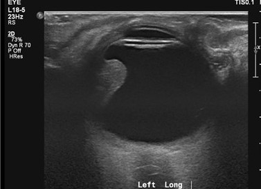
We have a several tests available to us to help us diagnose your condition and to help us decide on the best treatment. For choroidal melanoma, the most common type of eye cancer we see in our clinic, we routinely perform ultrasound scan of the eye in our radiology department. This is usually done before being seen in the eye clinic. Please see a list of investigations we may use below.
Ultrasound Scan
An Ultrasound scan is often used in pregnancy and uses sound waves to create an image of inside a part of the body. It is very safe and can look at organs in the abdomen, for example the liver, or of the eye. Either a doctor or a sonographer (health professional trained to perform ultrasound scans) will perform the scan. You may be asked to follow certain instructions before your scan- such as drinking a lot of water, fasting for a few hours etc- it is important you try and follow these instructions to get the best images possible. Cold gel is applied to the skin over the area they wish to scan.
The ultra-sound probe is gently placed on the skin and moved over the gel. If the eye is being scanned, the ultrasound probe is gently applied to the surface of the eyelid. The ultrasound images are then collected and analysed. Ultrasound scans of the body may take between 15-45minutes to perform. Ultrasound of the eye, however, is much quicker and will sometimes be performed in the eye clinic. Results of the eye USS will be given on the same day. Results of liver ultrasound scans, if carried out your local hospital, will be discussed at your next eye clinic appointment.
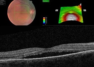
Optical Coherence Tomography (OCT)
This is a large camera, which creates 3D pictures of the back of the eye. After having your pupils dilated with drops, the scan takes minutes to perform. This helps us check for fluid leaking at the back of the eye from abnormal blood vessels or from a tumour. It can be repeated at each visit, and helps us pick up changes at the back of the eye.
CT Scan
CT scans are also known as CAT scans. They use several X-ray images to create a detailed scan of the body. When you arrive at the department you will have a short safety questionnaire to complete; due to radiation CT scans are not usually recommended in women who are pregnant. Sometimes contrast dye is injected into the vein during the scan to help get more detailed images.
The CT scanner itself is a large circular machine, which you lie flat in. You will be asked to stay still and breathe normally. The x-ray parts move inside the big circular ring creating the images. During the scan, you will be able to communicate with the radiographer in the next room through an intercom system. The scan takes around 10 to 20 minutes to do. After the scan, the images are analysed and the results will be given to you at your next clinic appointment.
MRI Scan
Add/copy info from UmbMRI stands for magnetic resonance imaging. Instead of X-ray radiation, It uses magnets and radio waves to produce images of inside the body. It gives very fine detailed pictures of the eye, the optic nerve, and the brain. When you arrive at the department you will have a short safety questionnaire to complete; if you have any metal inside the body or are claustrophobic you may not be able to get the MRI scan. Sometimes contrast dye is injected into the vein during the scan to help get more detailed images.
The MRI scanner is a large circular tube, which you lay down flat in. The radiographer then leaves the room to operate the scanner from the next room. You will be able to communicate with them however through an intercom system. During the scan you will be asked to lie still and to breathe normally. You may hear loud tapping noises during the scan- this is normal. You will be given earplugs or headphones to wear so you can listen to music. The scan takes 30 minutes to an 1 hour to complete. After the scan, the images are analysed and the results will be given to you at your next clinic appointment.
Chest X-Ray
X-rays are used to produce pictures of the heart, lungs and other parts of the body. This is quick to perform in the radiography department and is useful if we want to check for lung cancer.
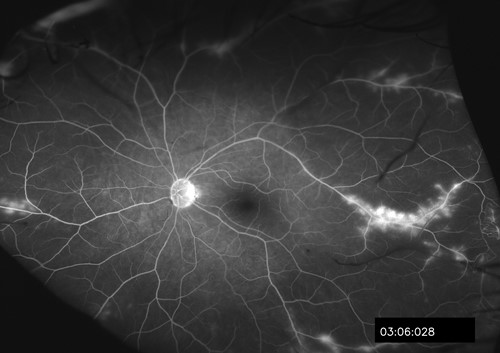
Fundus Fluorescein Angiography (FFA)
This is where a yellow dye is injection into a vein in the arm, which travels through the blood vessels to the back of the eye. As the dye reaches the back of the eye, several pictures are taken with a special camera. This gives us more information about the blood supply to suspicious areas at the back of the eye.
Indocyanide Green Angiography (ICG)
This is similar to fluorescein angiography, except a different dye is used. This looks more closely at the choroid at the back of the eye, which is the layer deeper to the retina. It is therefore useful at detecting abnormalities in the blood vessels in the choroid.
Patients and staff work hard to organise events to raise money for the Scottish Ocular Oncology Service. As a tribute to the efforts made to raise money for our service, highlights from past and future events are included in this section.
24 Hour Bowl-a-thon
The 24 hour Bowl-a-thon organised by one of our patients took place at the Renton Bowling club, Dunbartonshire, on the 8th/9th July 2017. This was a hugely successful event raising over £3,500 for the Scottish Ocular Oncology Service.
West Highland Way Fundraiser
On Easter Monday 2018 one of our patients Elaine and her sister, cousin, son, daughter, friend and niece walked part of the west highland way from Crainlarich to Bridge of Orkey.
From their heroic efforts they managed to raise a fantastic £1,454 for Scottish Ocular Oncology Service. Well done!
The Orbit is the bony socket that contains and protects the eye. It also contains blood vessels, ligaments, muscles and nerves that help the eye move and see. Fat cushions these structures in the orbit. In the top outer corner of orbit sits the lacrimal gland. This produces tears helping keep the eye moist.
Several different tumours can develop within the orbital cavity. Some are benign, some are malignant. This can cause double vision, decreased vision, and make the eye stick out (proptosis- picture above)
Please see the different orbital tumours below.
Cavernous Haemangioma
a) What is cavernous haemangioma?
This is where abnormally dilated blood vessels behind the eye collect together and form a tumour. This is the most common benign orbital tumour in adults. It occurs more often in females and can develop anywhere in the orbit. More commonly, however, it develops beside the muscles that move the eye. Although this tumour is benign, it can grow slowly behind the eye and cause damage.
b) What are the symptoms of cavernous haemangioma?
There may be no symptoms. If the cavernous haemangioma continues to grow, however, it can cause the following:
• Proptosis (Eye sticking out)
• Double vision
• Blurring of vision
c) Will I need any tests?
We may take pictures of the eye and consider the following tests:
• CT scan
• MRI scan (picture above)
This can help us measure the size of tumour and where it is. This is useful if we are planning surgery or to check if the nerve is being squashed. Cavernous haemangioma on MRI scan.
d) What is the treatment of cavernous haemangioma?
If not causing problems we may just observe routinely in the clinic. If, however, it is growing and causing problems, surgery is the best treatment. Unfortunately bleeding, infection or damage to nerves and muscles of the eye can occur during surgery. The risk of this depends on the tumour location and size.
Lacrimal Gland Pleomorphic Adenoma
a) What is pleomorphic adenoma?
This is the most common benign tumour of the lacrimal gland. It is also known as benign mixed cell tumour. It is formed from the cells and ducts that produce tears in the lacrimal gland. They also commonly occur in the salivary glands. Although labelled as benign, very rarely pleomorphic adenoma can turn into malignant tumours.
b) What are the symptoms of lacrimal gland pleomorphic adenoma?
This causes a painless lump in the lacrimal gland and sometimes swelling to the top eyelid. This can slowly cause the eye to stick out (proptosis – picture above). It can also push the eye down and inwards. This may cause double vision and decreased the vision.
c) Will I need any tests?
Different types of scans can be used to help diagnose this benign tumour. It is also useful if surgery is being planned. Scans may include:
• CT scan
• MRI scan
• US scan
d) What is the treatment for lacrimal gland pleomorphic adenoma?
Removal with surgery may be required. We aim to remove the whole tumour without cutting into the tumour – this may cause it to spread and re-grow at a later stage. For this reason biopsy we do not take a biopsy before surgery.
Lacrimal Gland Carcinoma
a) What is lacrimal gland carcinoma?
This is a rare malignant tumour of the lacrimal gland. The commonest malignant lacrimal gland carcinoma is called adenoid cystic carcinoma. This tumour can grow quickly and spread to the bones and the nerves around the eye.
b) What are the symptoms of lacrimal gland carcinoma?
This tumour can grow quickly and cause swelling of the top eyelid. The eye may stick out (proptosis- picture above) or be pushed downwards and inwards. This can cause double vision and blurring. If the tumour spreads into the bone and the nerves this can cause pain or numbness over the face.
c) What are the risks of getting lacrimal gland carcinoma?
The following increase your risk of lacrimal gland carcinoma:
• Age – it is more common in your 30s.
• Previous radiation treatment to the face,
• Previous removal of a benign lacrimal gland pleomorphic adenoma.
d) Will I need any tests?
We may perform the following scans:
• CT scan
• MRI scan
• Ultrasound scan
This helps us measure the size of the tumour, position, and check if there is any spread into the bone. Biopsy of the tumour is sometimes performed to confirm the diagnosis in the laboratory.
e) How is lacrimal gland carcinoma treated?
Surgery and radiotherapy is usually required. Surgery may include removal of the tumour and tissue around the lacrimal gland. If the tumour has spread out of the lacrimal gland then exenteration may be required. Unfortunately treatment is rarely curative, however, our medical oncology team will guide us on the best treatment for you.
Optic Nerve Glioma
a) What is optic nerve glioma?
Optic nerve glioma is a slow growing tumour developing within the optic nerve. It is more common under the age of 20. Another name for this tumour is a pilocytic astrocytoma. Patients who get this tumour commonly have an under lying medical condition called neurofibromatosis type 1. This is a genetic condition that makes you more likely to grow tumours along your nerves.
b) What are the symptoms of optic nerve glioma?
Since this tumour is slow growing, it usually causes a gradual decrease in vision in the affected eye. The tumour can push the back of the eye causing the eye to stick out (proptosis- picture above). Rarely, the vision may quickly deteriorate. This may be due to bleeding into the tumour, causing it to squash the nerve or the blood vessels to the eye. Sometimes the tumour can spread backwards into the brain. If this happens then headaches (worse in the morning), nausea and vomiting may occur.
c) Will I need any tests?
If optic nerve glioma is suspected then scanning the orbit and the brain is likely to be required. This may include:
• MRI scan
• CT scan
• US scan
If neurofibromatosis is suspected then genetic testing may be performed. A full body scan may also be useful to look for other tumours else where in the body. If the diagnosis is uncertain we may decide to take biopsy.
d) What is the treatment for optic nerve glioma?
If not causing any problems then treatment may not be required. If, however, the tumour is growing and causing forward displacement of the eye or decreasing the vision, then surgery is recommended. Unfortunately, because the tumour is on the optic nerve, removing this may cause damage to the vision. Radiotherapy and chemotherapy may also be used if the cancer has spread to the brain. Our medical oncology team will guide us on the best treatment for your case.
e) How effective is the treatment?
Treatment is effective in slow growing optic nerve gliomas. These are more common in children. In adults, however, aggressive fast growing optic nerve gliomas are more common and are more likely to spread back into the brain and threaten life. These are more difficult to treat.
Optic Nerve Sheath Meningioma
a) What is optic nerve sheath meningioma?
Wrapped around the optic nerve is a sheath. This is similar to the insulation layer of a wire. Along this sheath, a benign tumour called a meningioma can develop. This more commonly occurs between 30 and 60 years of life. It is 4 times more likely to occur in woman. This tumour can also develop in the protective layer of the brain and the spinal cord. Meningiomas may grow fast or slow.
b) What are the symptoms of optic nerve sheath meningioma?
The faster the tumour is growing then the quicker the symptoms will develop. This may include:
• Poor vision
• Proptosis (eye sticking out)
• Double vision .
c) Will I need any tests?
If meningioma is suspected, scanning of the orbit and the brain is usually carried out. Scans may include:
• MRI scan (picture above)
• CT scan
• US scan
Scans may be repeated at a later date to see if the meningioma is growing. If the diagnosis is uncertain we may decide to take a biopsy.
d) What is the treatment of optic nerve meningioma?
Treatment may not be required if the tumour is slow growing and not causing symptoms. This is more common in middle aged patients. In younger patients, however, the tumour is more likely to be aggressive and fast growing. If this is the case, surgery is required. Unfortunately surgery may damage the nerve and cause poor vision. Sometimes radiotherapy is given after surgery to reduce the risk of it coming back later in life. Unfortunately, radiotherapy may also damage the vision.
Lymphoma
a) What is lymphoma?
In lymphoma, the white cells that usually fight infection are “out of control”, keep dividing, and do not die. These abnormal cells collect and grow in the lymph nodes. They can also develop elsewhere in the body, including behind the eye. There are two different types of lymphoma – hodgkins lymphoma (20% of cases) and non-hodgkins lymphoma (80% of cases). Hodgkins lymphoma is the easiest to treat, however, treatments for both types of lymphoma are very effective and can be cured in most cases.
b) What are the symptoms of orbital lymphoma?
This may cause:
• Proptosis (eye sticking out- picture above)
• Double vision
• Decreased vision
c) Will I need any tests?
A biopsy may be required to confirm the diagnosis. Scanning the eye, orbit, and rest of the body is important to make sure the lymphoma is not affecting anywhere else. This may include:
• CT scan
• MRI scan
• US scan
This helps us decide which treatment is needed.
d) What are the treatments for orbital lymphoma?
Treatments include:
• Radiotherapy
• Surgery
• Chemotherapy
Treatments vary patient to patient, however, discussion with our medical oncologist and pathologist will help us choose the best treatment for you.
Orbital Metastases
a) What are orbital metastases?
Tumours starting elsewhere in the body can spread behind the eye in the orbit. This is rare, however, if a tumour is first found in the orbit then cancer elsewhere may have to be considered and looked for. Common places for cancer to grow first before spreading to the eye include the breasts, lungs, prostate, skin, bowel and kidneys.
b) What are the symptoms of orbital metastasis?
Orbital metastases may cause:
• Proptosis (eye sticking out- picture above)
• Double vision
• Decreased vision
In certain tumours, enophthalmos can occur; this is where the eye moves backwards into the orbit making the eye appear smaller. If cancer elsewhere is present then some of the following symptoms may be experienced:
• Shortness of breath
• Long standing cough
• Coughing up blood
• Breast lump
• Difficulty passing urine
• Blood in urine or stools
• Change in bowel habit
c) What tests will I get?
Scanning the eye and elsewhere in the body depending on symptoms experienced. Scans may include:
• CT scan
• MRI scan
• PET CT scan
• Chest X-ray
• Blood tests
If we are unsure of the diagnosis, an orbital biopsy may be carried out. This, however, will be discussed further in clinic.
d) What is the treatment for orbital metastasis?
Treatments may include:
• Radiotherapy
• Chemotherapy
• Surgery
The main goal is to preserve vision and keep the eye comfortable. Our medical oncologist will help us choose the best treatment for you.
Eyelids are thin folds of skin that cover and protect the eye. When blinking, the eyelid spread tears over the eye keeping it moist. There are several different types of cancer can develop on the skin of the eyelid.
The large majority of these can be managed at your local hospital. If there are concerns, however, of the cancer spreading behind the eye or else where in the body then we may be asked to help.
Please see the different eyelid tumours below.
Basal Cell Carcinoma
a) What is basal cell carcinoma?
Basal cell carcinoma (BCC) is the commonest type of skin cancer. The number of BCCs diagnosed is increasing every year due to the hotter summers in the UK and increasing foreign travel. These can grow on the skin of your eyelid. Due to their appearance they are often called ‘rodent ulcers’. They can grow along the eyelid, and if left untreated, can spread into or behind the eye. It is very rare for BCCs to spread around the body and can be cured if removed.
b) What are the symptoms of basal cell carcinoma?
Usually they are painless. They often look like a flat red mark, or have a pearly rim surrounding a crater. Sometimes they can bleed and scab over.
c) Risk Factors Risk factors for this condition include:
• Sun damage
• Fair skin
• Previous basal cell carcinomas
• Open wounds that resist healing
• Exposure to ionising radiation
• Chronic inflammatory skin conditions
d) What tests will I need?
A biopsy may be taken from the affected area. If it is suspected that the BCC has started to spread into the eye or behind the eye, scans may have to be performed. These may include:
• CT scan
• MRI scan
• Ultrasound scan
e) How are basal cell carcinomas treated?
Treatments include:
• Surgery (surgical excision)
• Radiotherapy
• Chemotherapy cream (5 Fluorouracil)
After surgery, the eyelid is reconstructed to make the eyelid appear and function as normal as possible. This may be carried out as a separate operation.
Squamous Cell Carcinoma
a) What is squamous cell carcinoma?
Squamous cell carcinoma (SCC) is the second most common skin cancer behind basal cell carcinoma. This is more aggressive, however, and can spread to other parts of the body. It can be tricky to diagnose on clinical appearance alone. The number of SCCs diagnosed in the world, and in the UK, is increasing each year. They are more common in Caucasians, in the elderly and in the male population.
b) What are the risks of squamous cell carcinoma spreading around the body?
Factors that increase the risk of metastases include:
• Large SCC greater than 2cm in diameter or deeper than 4mm
• SCC after radiation treatment
• SCC recurrence after surgical resection
• Long standing ulceration from Bowen’s disease (skin condition)
• Patients with a poor immune system from certain treatments or diseases
If the cancer has started to spread swelling of the lymph nodes around your ear or under your chin may occur.
c) What are the symptoms of squamous cell carcinoma?
They often appear as a non-healing ulcer with hard raised edges. There may be redness, crusting or bleeding. They commonly occur on the lower eyelid. Decreased vision may occur if the cancer spreads behind the eye.
d) What are the risks of getting squamous cell carcinoma?
Risk factors for this condition include:
• Sun damage
• Fair skin
• Pre-malignant conditions such as Bowens disease, actinic keratosis or keratoacanthomas.
• Poor immune system (e.g. HIV)
e) What tests will I need?
A biopsy will likely be taken. If there is high suspicion of SCC, the abnormal area of skin may be completely removed in theatre. If it is suspected that the SCC has started to spread into the eye or behind the eye we may arrange the following scans:
• CT scan
• MRI scan
• Ultrasound scan
f) Management Treatments include:
• Surgery (surgical excision)
• Radiotherapy
• Chemotherapy cream (5 Fluorouracil)
After surgery, the eyelid is reconstructed to make the eyelid appear and function as normal as possible. This may be carried out as a separate operation.
Sebaceous Gland Carcinoma
a) What is sebaceous gland carcinoma?
Sebaceous gland carcinoma is a rare and aggressive type of skin cancer. They grow from sebaceous glands, which normally produce our oil for our skin. These glands are also found in the eyelids. This can be tricky to diagnose as it is often mistaken for a chalazion or blepharitis – two common benign eyelid conditions. If there is a delay in treatment then the tumour may spread to other parts of the body including the liver, lungs, brain and bones. This can occur in up to a quarter of patients.
b) What are the symptoms of sebaceous gland carcinoma?
This commonly looks like a small, red or yellow, firm lump on the eyelid. This may gradually increase in size and cause irritation to the eye.
c) What are the risks of getting sebaceous gland carcinoma?
The following increase your risk of getting sebaceous gland carcinoma:
• Getting benign adenomas (non cancerous lumps) of the skin.
• Exposure to radiation e.g. radiotherapy to the face
• Asian ethnicity Spreading to other areas of the body unfortunately occurs in around 1 out of 5 cases.
d) Will I need any tests?
After taking a picture we will likely take a biopsy of the suspicious area and send it to the laboratory for testing. If we suspect the cancer has spread behind the eye or to other parts of the body we may organise the following scans:
• Chest X-ray
• CT scan
• MRI scan
e) What is the treatment for sebaceous gland carcinoma?
Treatments include:
• Surgery (surgical excision)
• Radiotherapy
• Chemotherapy cream (5 Fluorouracil)
After surgery, the eyelid is reconstructed to make the eyelid appear and function as normal as possible. This may be carried out as a separate operation Biopsy or removal of the lymph nodes may have to be performed if we suspect is has spread from the eyelid to the lymph nodes. Unfortunately the tumour can grow back after treatment.
Eyelid Melanoma
a) What is melanoma?
Melanoma is a serious type of skin cancer which can affect the eyelids. It grows from cells called melanocytes, which normally produce pigment in our skin, hair, and eyes. This gives them a dark and pigmented appearance Eyelid melanoma, although less common than other skin tumours, is dangerous because it can spread around the body if not treated early.
b) What are the symptoms of melanoma?
The following skin changes may be due to melanoma:
• Increased pigmentation or darkening of the skin
• A new mole
• A change in an existing mole
These are more likely to have an irregular shape, be more than one colour, and grow quickly.
c) What are the risks of getting eyelid melanoma?
The following increase your risk of getting eyelid melanoma:
• Pale skin
• Large numbers of moles or freckles
• Red or blond hair
• Blue eyes
• Poor immune system (e.g. HIV)
Melanoma can be prevented with good skin care when travelling abroad or in sunny climates. Careful monitoring of pigmented areas of skin is key to detecting skin melanoma early.
d) Will I need any tests?
We will likely take photographs to monitor changes. If the diagnosis is uncertain we may take remove all of the abnormal area and send it to the laboratory for where a diagnosis can be made . If it is suspected that the melanoma has started to spread to other parts of the body then we may organise the following scans:
• CT scan
• MRI scan
Blood tests, including liver function tests, may be carried out if spread to the liver is suspected. Liver ultrasounds carried out every year to monitor for spread at your local hospital.
e) What is the treatment for eyelid melanoma?
Treatments include:
• Surgery
• Radiotherapy
• Chemotherapy
If radiotherapy or chemotherapy is needed, our medical oncologist and pathologist will help guide our treatment.
f) What happens if the eye melanoma spreads to different parts of the body?
This can be scary and upsetting. If spread of disease has been picked up we involve other health professionals at our MDT (multidisciplinary team) meeting. Our clinical team consists of a radiologist, pathologist, and a medical oncologist. Collectively we will decide on the rights tests and treatments for you as an individual. Accepting and coming to terms with the diagnosis can be challenging. For this reason, we have specialist ophthalmic nursing staff in the clinic who are here to council and help you through this difficult time.
This sections looks at the different types of tumours that affect the conjunctiva (“the skin” of the eye, which helps protect us from infections).
Please see the conjunctival tumours below.
Ocular Surface Squamous Neoplasia
a) What s ocular surface squamous neoplasia?
Ocular surface squamous neoplasia is pre-cancerous or cancerous changes to the surface of the conjunctiva. Pre-cancerous changes can cause a condition called conjunctival intraepithelial neoplasia. This condition can turn into squamous cell carcinoma, which is a sinister tumour. This can spread onto the cornea causing decrease in vision. It usually affects one eye and is caused by sun damage.
b) What are the symptoms of ocular surface squamous neoplasia?
A fleshy pale lump is usually noticed on the surface of the eye next to the cornea. It may grow onto the cornea and, if large, can cause:
• Grittiness to the eye
• Watering
• Blurring of vision
Sometimes it can grow form the conjunctiva onto the eye lid.
c) What are the risks of getting ocular surface squamous neoplasia?
Risk factors for this condition include:
• Sun damage
• Fair skin
• Older age
• Conditions decreasing the immune system (e.g. HIV)
d) Will I need any tests?
In the clinic we may take pictures of the front of the eye. It is difficult to tell apart conjunctival intraepithelial neoplasia (pre-cancerous) from squamous cell carcinoma (cancerous). If the abnormal area is removed with surgery then it is sent to the laboratory to help confirm the diagnosis.
e) What is the treatment for ocular surface squamous neoplasia?
The main treatment is surgery (surgical excision). We may, however, use freezing treatment (cryotherapy) and Mitomycin C (MMC) eye drops as well. MMC can, however, irritate the eye. To treat this we may provide you with lubricant or steroid eye drops.
Primary Acquired Melanosis
a) What is primary acquired melanosis?
Primary Acquired melanosis (PAM) is newly formed brown pigmentation of the conjunctiva. People with white skin are more likely to get this. It usually affects one eye only. There are two different types of PAM: ‘PAM without atypia’ and ‘PAM with atypia.’ PAM without atypia is benign with no risk of transformation into melanoma. PAM with atypia, however, can turn into melanoma. Around 3 out of 4 conjunctival melanomas come from PAM with atypia. If you have dark skin, brown pigmentation of the conjunctiva from birth is very common and usually affects both eyes. This is known as conjunctival epithelial melanosis and is not likely to progress to melanoma.
b) What are the symptoms of primary acquired melanosis?
Uneven, painless, newly formed brown pigmentation is usually noted on the white of the eye. If the pigment is growing quickly or changing colour then it is more likely to be a conjunctival melanoma.
c) Am I likely to get primary acquired melanosis?
If you are middle aged and fair-skinned you are more likely to get primary acquired melanosis.
d) Will I need any tests to exclude melanoma?
If it is felt to be PAM without atypia we may take pictures in the clinic to help us monitor change. Sometimes a biopsy of the pigmented area, however, is needed to exclude cancer. When doing so we try to remove all of the pigmented area. The following features make us more likely to biopsy:
• Larger than 5mm
• Increasing in size
• Has thickened the conjunctiva
• Has a distinct nodule arising within it (pigmented or non-pigmented raised area)
• Has feeder vessels (blood vessels running into the pigmented area)
• Involving the cornea
• Involving the conjunctiva under the eyelids
• Previous melanoma
e) Is treatment needed for primary acquired melanosis?
PAM without atypia can be monitored in the clinic. The main treatment for PAM with atypia is surgery (surgical excision) with or without freezing therapy (cryotherapy) and Mitomycin C (MMC).
Conjunctival Melanoma
a) What is conjunctival melanoma?
Melanomas are tumours which come from pigmented cells in our body. As well as growing on the skin, they can grow on the conjunctiva of the eye. Conjunctival melanoma accounts for 2% of eye cancers. It arises most commonly form primary acquired melanosis, or from a naevus (freckle) already present on the eye. Least commonly it arises on its own; this is known as primary conjunctival melanoma. This cancer can spread to other organs in the body, most commonly, the liver.
b) What does conjunctival melanoma look like?
Conjunctival melanoma looks like an uneven, raised, grey or pink lump growing on white conjunctiva or from a pigmented area on the conjunctiva. This may cause irritation and watering from the eye.
c) What are the risks of getting conjunctival melanoma?
The following increase your risk of developing conjunctival melanoma:
• Having pale skin
• Red or blond hair
• Blue eyes
• Over 50 years of age
• Large number of moles or freckles
• Poor immune system (e.g. HIV)
d) How likely is the conjunctival melanoma to spread around the body?
There are some features that increase the risk of the melanoma spreading. These include:
• Large tumours (large basal diameter, and tumour thickness of greater > 2mm)
• Nodular tumours (rounded and raised)
• Tumour involving the eyelids
• Tumour involving the corner of the eye near the nose (plica or caruncle)
If the tumour has grown on white conjunctiva instead of a pigmented conjunctiva, then the tumour may be more aggressive. Most common place is the liver. A liver ultrasound scan is arranged at your local hospital every year to exclude spread of disease.
e) Will I need any tests?
A biopsy may be required to confirm the diagnosis. If we decide to biopsy we usually remove the whole abnormal area. We may perform CT or MRI scan to make sure the melanoma has not spread to other parts of the body. If the tumour is greater than 2mm thick or growing in the corner of the eye we may need to consider doing a sentinel lymph node biopsy (SLNB). This is where a sample is taken from the glands that drain fluid away from the eye and analysed for tumour cells. These glands are found at the front of the ear and in the neck. This is the first place the tumour spreads to before metastasising around the body. This procedure is carried out in the Queen Elizabeth University Hospital in Glasgow with help from our maxilo-facial surgeon colleagues. If the SNLB detects cancer cells then the glands can be removed with surgery or treated with radiotherapy.
f) What is the treatment of conjunctival melanoma?
Surgery (surgical excision) is performed with or without:
• Cryotherapy
• Mitomycin C (MMC)
• Radiotherapy
• Chemotherapy
Sometimes the tumour is too big or has spread behind the eye. In these situations enucleation may be the best option. If there is involvement of the eyelids or of tissue behind the eye, we may have to consider exenteration. If there is spread of disease to the lymph nodes in the neck, we may have to remove these or treat with radiotherapy.
g) Can the conjunctival melanoma come back after treatment?
Unfortunately the answer to this is yes. Even if the tumour is completely removed there is always a chance of the tumour re-growing in the same place or in other parts of the body. For this reason, we monitor in the clinic after treatment.
Lymphoma
a) What is lymphoma?
Lymphoma is a condition where the white cells that usually fight infection are “out of control”, keep dividing, and do not die. These abnormal cells can collect and grow in the lymph nodes. They can, however, grow elsewhere in the body including the conjunctiva. Conjunctival lymphomas are mostly of non-Hodgkin’s B-cell type. Follicular lymphoma is the next most common lymphoma. The lymphoma may just affect the conjunctiva or can affect other areas in the body as well.
b) What are the symptoms of conjunctival lymphoma?
Conjunctival lymphoma looks like a large “salmon pink” growth on the conjunctiva. This may cause grittiness, watering, or irritation to the eye.
c) Will I need any tests?
A biopsy is usually required to confirm the diagnosis. If we suspect the lymphoma is behind the eye or affecting else where in the body we may organise:
• Ultrasound scan
• CT scan
• MRI scan
d) What is the treatment for lymphoma?
After biopsy confirms the diagnosis of lymphoma, we may treat with:
• Radiotherapy
• Chemotherapy
Our choice of treatment is be guided by the haematologist or medical oncologist.