We have a several tests available to us to help us diagnose your condition and to help us decide on the best treatment. For choroidal melanoma, the most common type of eye cancer we see in our clinic, we routinely perform ultrasound scan of the eye in our radiology department. This is usually done before being seen in the eye clinic. Please see a list of investigations we may use below.
Ultrasound Scan
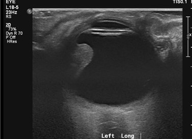
An Ultrasound scan is often used in pregnancy and uses sound waves to create an image of inside a part of the body. It is very safe and can look at organs in the abdomen, for example the liver, or of the eye. Either a doctor or a sonographer (health professional trained to perform ultrasound scans) will perform the scan. You may be asked to follow certain instructions before your scan- such as drinking a lot of water, fasting for a few hours etc- it is important you try and follow these instructions to get the best images possible. Cold gel is applied to the skin over the area they wish to scan.
The ultra-sound probe is gently placed on the skin and moved over the gel. If the eye is being scanned, the ultrasound probe is gently applied to the surface of the eyelid. The ultrasound images are then collected and analysed. Ultrasound scans of the body may take between 15-45minutes to perform. Ultrasound of the eye, however, is much quicker and will sometimes be performed in the eye clinic. Results of the eye USS will be given on the same day. Results of liver ultrasound scans, if carried out your local hospital, will be discussed at your next eye clinic appointment.
Optical Coherence Tomography (OCT)
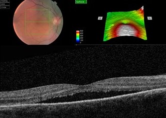
This is a large camera, which creates 3D pictures of the back of the eye. After having your pupils dilated with drops, the scan takes minutes to perform. This helps us check for fluid leaking at the back of the eye from abnormal blood vessels or from a tumour. It can be repeated at each visit, and helps us pick up changes at the back of the eye.
CT Scan
CT scans are also known as CAT scans. They use several X-ray images to create a detailed scan of the body. When you arrive at the department you will have a short safety questionnaire to complete; due to radiation CT scans are not usually recommended in women who are pregnant. Sometimes contrast dye is injected into the vein during the scan to help get more detailed images.
The CT scanner itself is a large circular machine, which you lie flat in. You will be asked to stay still and breathe normally. The x-ray parts move inside the big circular ring creating the images. During the scan, you will be able to communicate with the radiographer in the next room through an intercom system. The scan takes around 10 to 20 minutes to do. After the scan, the images are analysed and the results will be given to you at your next clinic appointment.
MRI Scan
Add/copy info from UmbMRI stands for magnetic resonance imaging. Instead of X-ray radiation, It uses magnets and radio waves to produce images of inside the body. It gives very fine detailed pictures of the eye, the optic nerve, and the brain. When you arrive at the department you will have a short safety questionnaire to complete; if you have any metal inside the body or are claustrophobic you may not be able to get the MRI scan. Sometimes contrast dye is injected into the vein during the scan to help get more detailed images.
The MRI scanner is a large circular tube, which you lay down flat in. The radiographer then leaves the room to operate the scanner from the next room. You will be able to communicate with them however through an intercom system. During the scan you will be asked to lie still and to breathe normally. You may hear loud tapping noises during the scan- this is normal. You will be given earplugs or headphones to wear so you can listen to music. The scan takes 30 minutes to an 1 hour to complete. After the scan, the images are analysed and the results will be given to you at your next clinic appointment.
Chest X-Ray
X-rays are used to produce pictures of the heart, lungs and other parts of the body. This is quick to perform in the radiography department and is useful if we want to check for lung cancer.
Fundus Fluorescein Angiography (FFA)
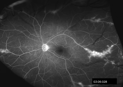
This is where a yellow dye is injection into a vein in the arm, which travels through the blood vessels to the back of the eye. As the dye reaches the back of the eye, several pictures are taken with a special camera. This gives us more information about the blood supply to suspicious areas at the back of the eye.
Indocyanide Green Angiography (ICG)
This is similar to fluorescein angiography, except a different dye is used. This looks more closely at the choroid at the back of the eye, which is the layer deeper to the retina. It is therefore useful at detecting abnormalities in the blood vessels in the choroid.